Yes, dental X-rays are safe for most people, including children, because they use a very low dose of radiation. A set of 4 bitewing X-rays with modern digital sensors delivers roughly the same radiation you’d get from about 1 day of normal background exposure, or a short flight. Concern about radiation is common and reasonable, but for routine dental imaging the risk is very small, and your dental clinic should only take X-rays when they’re likely to change your care.
Most people are surprised to learn how much a dentist can’t see with their eyes alone. Cavities between teeth, infections at the root tip, bone loss from gum disease, and impacted wisdom teeth are often invisible until an X-ray shows them. That’s why X-rays are part of regular dental hygiene and checkup visits, at intervals based on your personal risk.
X-rays also guide treatment. Your dentist relies on them to plan a root canal, check that a filling sealed properly, and see whether a tooth has enough healthy root to support a crown.
The same goes for crowns and bridges, where X-rays confirm the fit at the edges and the health of the supporting teeth. If you’ve ever wondered why your dental clinic in Richmond Hill asks for X-rays every so often, the answer is that small problems show up there first.
Key facts
- Digital dental X-rays use up to 80 to 90% less radiation than old film X-rays.
- 4 bitewings: roughly 0.005 mSv, about 1 day of background radiation.
- A panoramic X-ray: roughly 0.01 to 0.02 mSv.
- Frequency should be based on your risk, not a fixed schedule for everyone.
What is a dental X-ray?
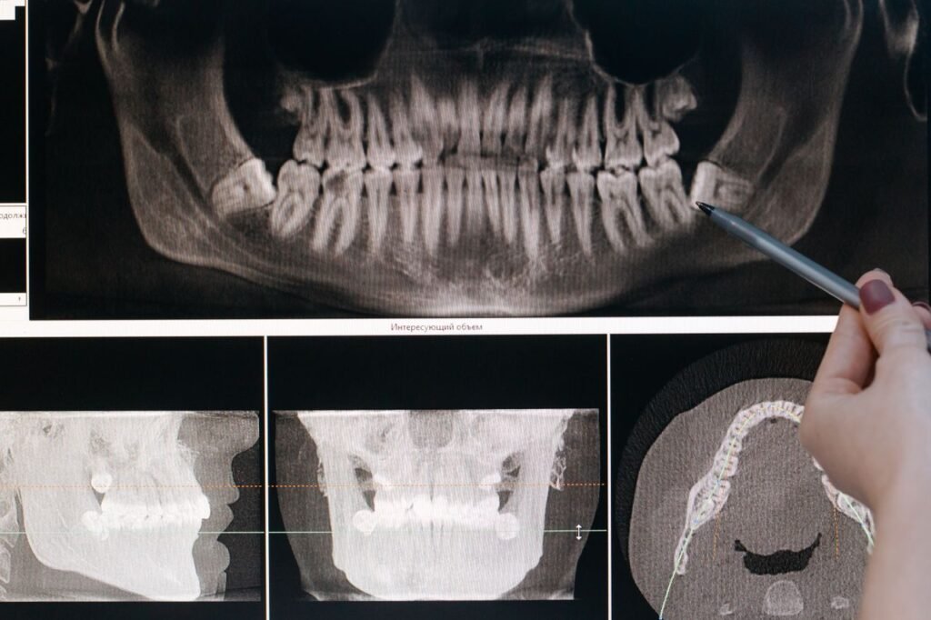
A dental X-ray (also called a dental radiograph) is an image made by passing a small, focused beam of X-ray energy through your teeth and jaw onto a sensor. Dense structures like enamel and bone block more of the beam and appear white. Softer areas, like decay or infection, let more through and appear darker.
What types of dental X-rays are there?
- Bitewing: shows the crowns of upper and lower back teeth together. Best for finding cavities between teeth and checking bone levels.
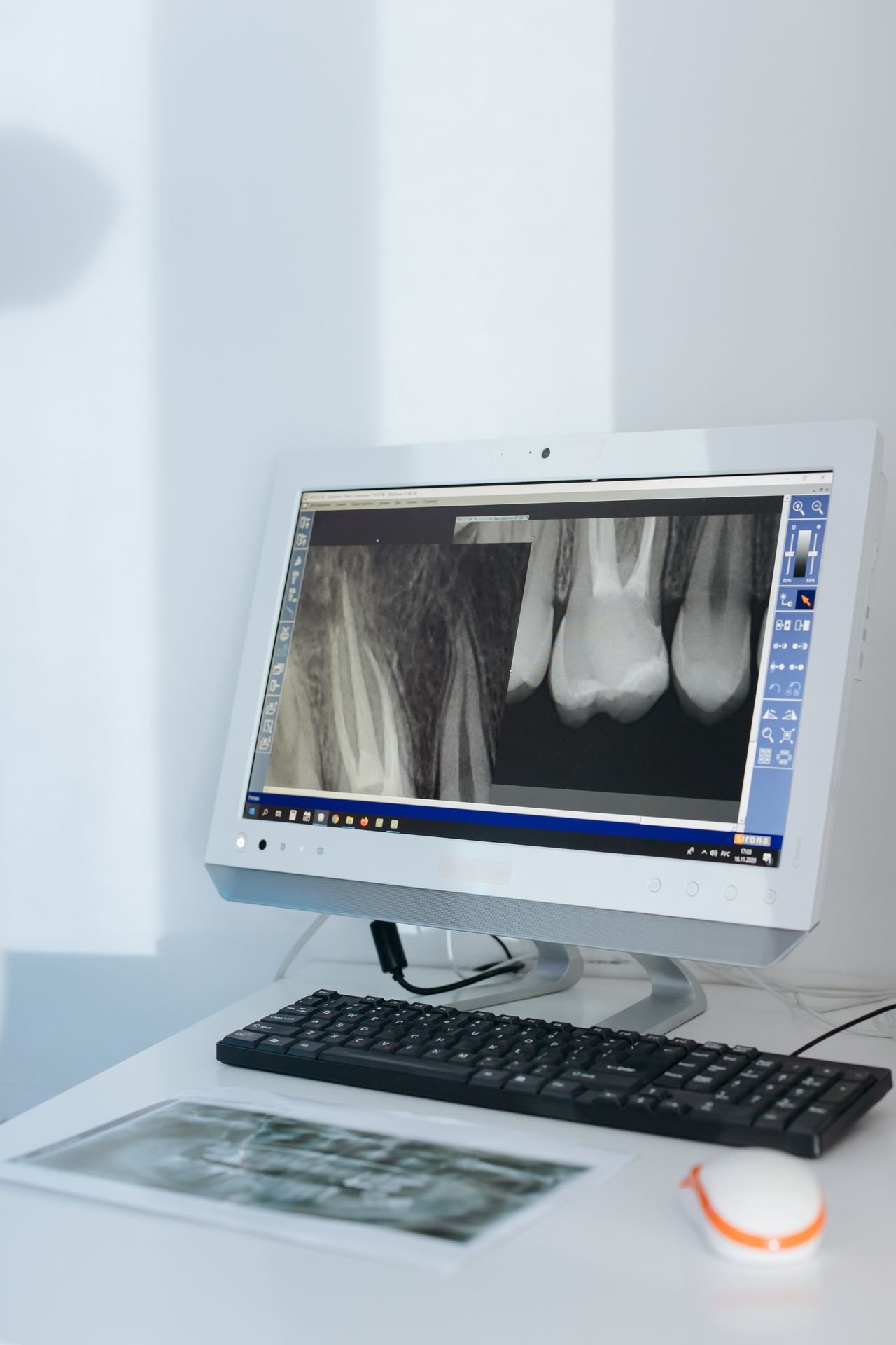
- Periapical (PA): shows a whole tooth from crown to root tip. Used for pain, infections, and root canal treatment.
- Panoramic: one wide image of the entire mouth, jaws, and sinuses. Good for wisdom teeth, orthodontic planning, and overall checks.
- Cone beam CT (CBCT): a 3D scan used for implant planning and complex cases. Higher dose, used only when needed.
How much radiation is in a dental X-ray, really?
Radiation dose is measured in millisieverts (mSv). The average person in Canada gets about 1.8 mSv per year from natural background radiation, like the ground, air, and food.
| Source | Approximate dose | Equal to about |
|---|---|---|
| 4 digital bitewings | 0.005 mSv | 1 day of background radiation |
| Panoramic X-ray | 0.01 to 0.02 mSv | 2 to 4 days |
| Cross-country flight (Toronto to Vancouver) | About 0.02 mSv | About 4 days |
| Chest X-ray | 0.1 mSv | About 10 days |
| Dental CBCT (small area) | 0.02 to 0.2 mSv | A few days to a few weeks |
| CT scan of the abdomen | 8 to 10 mSv | About 3 to 5 years |
These are averages. Exact doses depend on the machine, settings, and the area imaged.
Why do dentists still take X-rays if the risk is low?
Because “low” still isn’t zero, dental teams follow a principle called ALARA: As Low As Reasonably Achievable. Every X-ray should have a clear reason behind it, and the dose should be as small as possible.
The other side of the equation is what you’d miss without them. Studies suggest a visual exam alone misses a large share of cavities between back teeth. Early bone loss from gum disease, cysts, and infections at the root tip are often painless and invisible until they’re advanced.
How often should you get dental X-rays?
There’s no single schedule for everyone. Canadian and American guidelines recommend tailoring X-rays to each person’s risk.
| Patient | Typical bitewing interval |
|---|---|
| Child with high cavity risk | Every 6 to 12 months |
| Child with low cavity risk | Every 12 to 24 months |
| Adult with high cavity risk | Every 6 to 18 months |
| Adult with low cavity risk | Every 24 to 36 months |
| New patient | Often a baseline set, unless recent X-rays can be transferred |
If you’ve had X-rays at another office recently, ask for them to be sent over. That can save you from a repeat.
What can dental X-rays find that an exam can’t?
- Cavities between teeth and under old fillings
- Infections and abscesses at the tips of roots
- Bone loss from gum disease
- Impacted or sideways wisdom teeth
- Cracks or fractures in roots
- Cysts and, rarely, tumours in the jaw
- Extra or missing adult teeth in children
- How well a crown, bridge, or implant fits below the gumline
Many of these are painless for months or years. Finding them early usually means a smaller, simpler fix.
Are dental X-rays safe during pregnancy?
Dental X-rays are considered safe during pregnancy when they’re needed. The beam is aimed at the mouth, far from the abdomen, and the dose reaching the baby is extremely small. The American College of Obstetricians and Gynecologists and the Canadian Dental Association both support necessary dental imaging during pregnancy.
In practice, many dentists postpone routine, non-urgent X-rays until after delivery and take them only when there’s pain, swelling, or a suspected infection. Untreated dental infections pose a bigger risk to mother and baby than a properly shielded X-ray.
Always tell your dental team if you’re pregnant or might be.
Are dental X-rays safe for children?
Yes, with care. Children are more sensitive to radiation than adults because their cells are dividing faster, so dentists are more selective with them.
- Smaller sensors and lower settings are used for kids.
- X-rays are taken based on cavity risk and development, not on a fixed calendar.
- Panoramic images are often timed to check adult tooth development around ages 6 to 9 or before orthodontic treatment.
What about lead aprons and thyroid collars?
This is an area where guidance has shifted. Modern digital X-ray machines are tightly focused, and newer research shows lead aprons offer little added protection for the body during dental imaging. Some organizations, including the American Dental Association in 2023, no longer recommend routine aprons and collars.
Many patients still feel more comfortable with one, and that’s fine. Just ask. Thyroid collars can sometimes get in the way of the image and require a retake, so your dentist may suggest skipping it for certain views.
What happens during a dental X-ray?
- You’ll be asked about pregnancy and recent X-rays.
- A small sensor is placed in your mouth, or you stand at a panoramic machine.
- You hold still for a few seconds while the image is taken.
- The digital image appears on screen almost immediately.
- Your dentist reviews it with you and explains anything they find.
If you have a strong gag reflex, mention it. Smaller sensors, breathing techniques, and a panoramic image instead of individual films can help.
Myths about dental X-rays
“Dental X-rays cause cancer.”
Very high radiation doses can raise cancer risk. Dental X-ray doses are tiny by comparison, and the theoretical risk is extremely small. Some older studies linking dental X-rays to thyroid issues relied on people remembering past X-rays years later, which is unreliable, and involved older, higher-dose equipment.
“If nothing hurts, I don’t need X-rays.”
Most early cavities, bone loss, and root infections don’t hurt. By the time they do, treatment is usually bigger.
“Dentists take X-rays to bill more.”
Good practice ties every X-ray to your personal risk. You’re always allowed to ask why an X-ray is being taken and how it’ll change your care.
Real-life cases where X-rays mattered
The hidden cavity. A 26-year-old with no pain had bitewings after 3 years. They showed 2 cavities between molars. Small fillings fixed them. Left another year, one would likely have needed a root canal.
The silent infection. A man in his 50s had an old crown that felt fine. A periapical X-ray showed a dark area at the root tip. A root canal through the crown cleared the infection before it caused swelling.
The wisdom tooth. A teen’s panoramic image showed a lower wisdom tooth growing sideways into the molar in front. Planning removal early avoided damage to the neighbouring tooth.
Talking with your dentist about X-rays
You should never feel pressured. Good questions to ask:
- Why do I need this X-ray today?
- What are you looking for?
- Can you use X-rays from my previous dentist?
- What happens if I wait?
Richmond Hill Smile Centre is one of the best dental clinics in Richmond Hill, and the team uses digital imaging and explains each X-ray in plain language. If you’re looking for the best dentist in Richmond Hill for a careful, risk-based approach to checkups, it’s a good place to start.
The clinic is located at 10157 Yonge St Unit 101, Richmond Hill, ON L4C 1T6. Email info@richmondhillsmilecentre.ca to book an exam or to arrange the transfer of X-rays from a previous office. Care is always provided by licensed dental professionals at this dental office in Richmond Hill.
Frequently asked questions
Can I refuse dental X-rays?
Yes, it’s your choice. Your dentist will explain what they can and can’t diagnose without them, and some treatments, like root canals or implants, can’t be done safely without imaging.
Are digital X-rays safer than film?
Digital sensors need much less radiation to create an image, often up to 80 to 90% less than older film. They also produce images instantly and can be enlarged for a closer look.
Is a panoramic X-ray more dangerous than bitewings?
A panoramic X-ray uses a slightly higher dose than a set of bitewings but is still very low. It shows different things, so it’s used for different reasons.
How many dental X-rays are too many?
There isn’t a fixed number. What matters is that each X-ray has a clear purpose. If you’ve had many X-rays at different offices, sharing records helps avoid repeats.
Can X-rays show gum disease?
Yes. X-rays show the bone level around teeth, which helps diagnose and track periodontitis. Gum measurements during your exam complete the picture.
Conclusion
Dental X-rays use a very small amount of radiation and help catch problems an exam alone would miss.
How often you need them should depend on your personal risk, not a one-size-fits-all schedule.
Ask questions, share past records, and let your dentist explain how each image guides your care.